Nuclear Brain Imaging In Medical Practice In Japan
Dr. Jun Hatazawa, M.D., Ph.D.,
President of the AOFNMB, Japan
BASIS OF NUCLEAR BRAIN IMAGING
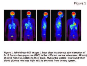
Brain is an organ by which we can perceive, exercise, learn, remember, think, behave and imagine among other tasks. To fulfill its functions, the brain exclusively metabolizes glucose to produce energy in the form of adenosine triphosphate (ATP) by oxidative glycolysis. Half of the ATP is used for the electrical activity of the neurons, and the other half is consumed for the synthesis and catalysis of brain tissue components. 18F fluoro-deoxy.glucose (18F FDG) PET imaging (figure 1) demonstrates very nicely the that that the human brain consumes more glucose than any other organ of the body. The human brain consumes approximately 100-150g of glucose per day.
Brain function is maintained by a continuous supply of glucose and oxygen by the cerebral circulation. The Cerebral Blood Flow (CBF) is tightly controlled to supply the brain with the
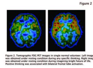
necessary amount of glucose and oxygen needed to perform its functions. Over the past few decades the scintigraphy mapping of the CBF has been used to determine and evaluate the activation of the different areas of the brain while performing various tasks such as speech, calculation, memory, and exercise (figure 2).
BRAIN STROKE
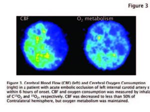
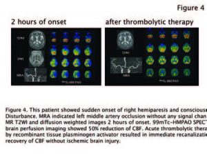
Brain is very sensitive to CBF reduction because of the need of a continuous and uninterrupted supply of glucose and oxygen. Brain perfusion imaging in nuclear medicine is now employed to detect CBF change in central nervous system diseases. Stroke is the most common neurological disease in adults and can be fatal. In acute ischemic stroke due to embolic occlusion of carotid artery or major cerebral arteries, CBF suddenly decreases to less than 50% of normal level. Patients complain of difficulty of motion, sensory loss, and finally consciousness disturbance. Even in such situation, the brain can still extract glucose and oxygen as much as possible from decreased CBF, and can survive during 6 hours of onset (figure 3). If the blood clot is resolved within 6 hours of onset, recirculation can minimize ischemic brain damage or rescue completely without any neurological symptoms (figure 4).
In chronic ischemic stroke due to atherosclerotic stenosis of major cerebral arteries, CBF is gradually decreased. Patients first complain of transient motion and/or sensory disturbance for several minutes once a month, followed by more frequent onset and longer duration. It indicates high probability of impending
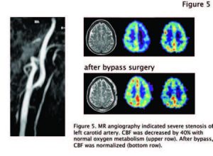
cerebral infarction with irreversible neurological deficits. Brain perfusion SPECT provides information on the severity of circulation disturbance before the onset of cerebral infarction and a guide for therapy strategy such as interventional reconstruction of stenotic artery, surgical removal of atheroma in thick arterial wall, or extra cranial-intracranial bypass surgery (Figure 5).
EPILEPSY
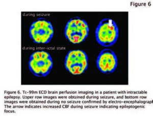
Epilepsy is one of the major neurological diseases in children. Most patients are treated with anti-epileptic medications, but around 10% of epileptic children needs further treatment such as surgical removal of epileptogenic foci. During epilepsy, epilep.togenic lesion shows abnormally increased CBF. Brain perfusion SPECT is employed to localize epileptogenic foci in patients with intractable epilepsy for surgical removal (Figure 6).
ALZHEIMER’S DISEASE
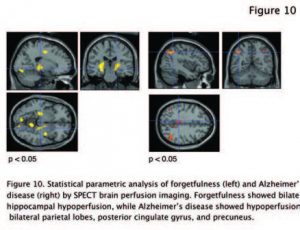
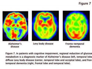
While aging, people experience forgetfulness. At the very early stage of Alzheimer’s disease (AD), patients complain of memory impairment. It is difficult to distinguish Alzheimer’s symptoms from physiological forgetfulness. Brain Perfusion Imaging in patients with cognitive impairment is now employed to categorize among AD, frontotemporal
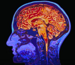
dementia, dementia with diffuse levy body disease, and other types (figure 7). Histopathological prediction of CNS diseases using PET/CT is now extending to Alzheimer’s disease where -amyloid and tau protein deposition is visualized.
PARKINSON’S DISEASE
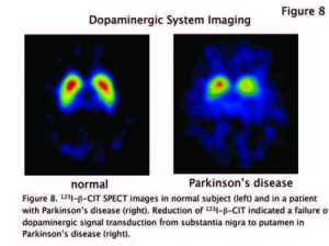
Brain functions require the sophisticated integration of neuronal signals among population of neurons. Communication among neurons is done by releasing chemical compounds called neurotransmitters released by a neuron and binding to another. Neuron to neuron communication failure happens due to neurotransmitter « dopamine » deficiency in Parkinson’s disease (figure 8), « acetylcholine » deficiency in Alzheimer’s disease, and « serotonin » deficiency in major depression. DatSCAN is a new SPECT tracer that enables the diagnostic of Parkinson’s disease (see Dr. Christian Scheiber article in this issue).
BRAIN TUMOR
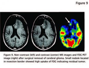
Brain tumor is one of the most dreadful diseases in adults and children. The therapeutic strategy is usually determined by histo.pathological classification according to World Health Organization grading. Biopsy of brain tumor is invasive because of transcranial tissue sampling. 18F-FDG or 11C-Methionine PET/CT is an alternative way to evaluate, non-invasively, the histopathological malignancy of cerebral gliomas non-invasively (figure 9).
NUCLEAR BRAIN IMAGING IN JAPAN
In Japan, one million and eighty thousand nuclear medicine studies are performed every year (National Survey of Nuclear Medicine Practice, Japan Radioisotope Association) using single photon isotopes on planar and SPECT cameras. Two hundred and fifty thousand of these studies, i.e. 22% are for brain imaging. Ischemic stroke, Parkinson’s syndrome, cognitive impairment, epilepsy, head injury (especially after traffic accident), and brain tumor are the major indications. The cost of the study is approximately $400~500 US. Our national insurance system covers 70% of the cost and patients pay about 30%.
BONE SCINTIGRAPHY (30%) AND MYOCARDIAL PERFUSION IMAGING (22%)
Compared to other countries and other nuclear medicine studies (bone scintigraphies and cardiac studies represent 30% and 22% respectively), in Japan is the availability and the widespread use of dedicated brain SPECT software such as eZIS (Dr. Hiroshi Matsuda and his colleagues) and iSSP (Dr. Satoshi Minoshima and his colleagues). In patients with cognitive impairment, hypo-perfusion is detected on a pixel-by-pixel and compared to a normal data base (Figure 10). It is objective, comprehensive, and reproducible for nuclear medicine physicians, referring doctors, and patients.
CONCLUSION
Nuclear brain imaging with SPECT and PET in Japan is a significant part of the nuclear medicine practice. It is applied to various brain disorders to detect physiological and metabolic abnormality before morphological changes become evident. The availability and use of performant and reliable analytical software is a significant factor for the success of brain imaging.
